Thursday, August 27, 2009

ProFibrix has raised $11 million

ProFibrix BV, a Dutch developer of products for hemostasis and regenerative medicine, has raised $11 million in Series B funding. Gilde Healthcare Partners led the round, and was joined by return backer Index Ventures.

PRESS RELEASE
ProFibrix B.V., a leader in the development of innovative products for hemostasis and regenerative medicine, today announced the successful closing of a US $11 million series B investment. The financing round was led by new investor Gilde Healthcare Partners, a leading European life sciences investor, who joins existing investor Index Ventures. Dirk Kersten from Gilde will join the company’s Supervisory Board.

Jaap Koopman, PhD, Chief Executive Officer, said: “We are very pleased to welcome Gilde Healthcare to our investor base and we appreciate the ongoing support and expertise that our investors can offer to our experienced team as we continue the development of ProFibrix. The proceeds of the financing will be applied to the ongoing clinical development of our lead product Fibrocaps(TM), and to the preclinical development of our pipeline products.”

Fibrocaps is based on a mixture of two essential blood clotting proteins, fibrinogen and thrombin, and is a unique dry powder topical tissue sealant that rapidly stops bleeding after or during surgery. Fibrocaps has major advantages over existing liquid tissue sealants: it is ready for immediate use, is stable at room temperature, highly effective and fast acting. Fibrocaps is currently in a Phase II clinical trial in the Netherlands with results expected end of 2009.

Dirk Kersten, investment manager at Gilde Healthcare Partners, commented: “Gilde Healthcare Partners invests in biopharmaceutical companies with strong technology platforms and experienced management. We believe ProFibrix’ breakthrough fibrinogen technology is well-positioned to play an important role in the highly attractive hemostasis and regenerative medicine markets.”

ProFibrix expects to submit an Investigational New Drug Application (IND) for Fibrocaps to the U.S. Food and Drug Administration in the first half of 2010, and to conduct a combined phase II/III pivotal study in various surgical indications.

Sea Worm Provides Model for Surgical Adhesive

Scientists at the University of Utah have uncovered a way to produce a synthetic adhesive based on a natural glue created by the sandcastle worm. This ocean worm builds its home by sticking pieces of sand and shells together, and the result is strong enough to withstand ocean tides. Researchers think this glue could have applications for bone repair in trauma cases, where fast setting time, ease of use and low risk of infection can benefit both the patient and the surgeon.

Russell Stewart, Ph.D., who led the Utah team, said his goal was to develop a water-based adhesive that remained insoluble in wet environments and was able to bond to wet objects. Curing time was another key issue for Stewart. The the team learned that pH changes cause the glue to set, a response that was copied for the synthetic version. The final result is "at least as strong as Super Glue" and is twice as strong as the sandcastle adhesive. So far, it has passed toxicity studies in cell cultures. In addition to its adhesive applications, Stewart is exploring how the glue could also be used to deliver antibiotics or materials that could aid in healing, like BMPs or other small molecules.

Surgical adhesives offer a number of benefits over fixation devices, among them more effective wound closure, less risk of leakage and infection and ease of handling and application. Similar to the sandcastle glue, many adhesives are derived from natural sources. For example, fibrin sealants are blood-derived, CryoLife's BioGlue is bovine-derived and some hemostatic products come from snake venom.

Thursday, August 20, 2009

Zymogenetics says rival’s product could be deadly

ZymoGenetics Inc. Thursday filed a petition with the U.S. Food and Drug Administration, saying that a rival drug company’s product could cause patients’ deaths and is asking the FDA to remove the drug from the market “in the interest of patient safety.”
The Seattle biotech (NASDAQ: ZGEN) filed a citizen petition, asking the FDA to remove King Pharmaceuticals Inc.’s Thrombin-JMI Thrombin topical (bovine origin) drug from the market because of “recent report of serious or fatal bleeding-related adverse events in surgical patients exposed to bovine (cattle-derived) thrombin.”
“Serious adverse events, including death, linked to bovine thrombin continue to be reported to the FDA. These adverse events are a serious, ongoing safety issue for patients undergoing surgery,” said Dr. George Rodgers, professor of medicine and pathology at the University of Utah, and medical director of the coagulation laboratory at ARUP Laboratories.
A spokesman for King said the Bristol, Tenn. company (NYSE: KG) had “no comment” on ZymoGenetics’ allegations.

Wednesday, August 19, 2009

Helixate FS Receives FDA Approval for Routine Prophylaxis in Children with Hemophilia A

CSL Behring announced today thatHelixate® FS, Antihemophilic Factor (Recombinant), has been approved by the U.S. Food and Drug Administration (FDA) for routine prophylaxis in children with hemophilia A who are 16 years old or younger and do not have pre-existing joint damage. This new indication was granted by the FDA after determining Helixate FS is safe and effective for routine prophylaxis, which can reduce the frequency of bleeding episodes and the risk of joint damage in children with hemophilia A.
“The FDA's approval of this additional indication for Helixate FS will significantly benefit the bleeding disorders community,” said Garrett E. Bergman, M.D. Senior Director of Medical Affairs, U.S. Commercial Operations at CSL Behring. “Prophylactic treatment will reduce the frequency of bleeding episodes in children with hemophilia A which, over time, will help prevent additional joint damage caused by bleeds.”
"Bleeding into joints, including the ankles, elbows and knees, is a concern for children with hemophilia A and their treatment providers," said Jerry S. Powell, M.D., Director of the Hemophilia Treatment Center at the University of California, Davis. "The FDA's approval of Helixate FS for routine prophylaxis is an innovation that will help our patient population effectively manage their condition. Modern treatment of hemophilia means preventing bleeding through routine prophylaxis, and we eagerly anticipate our patients reaching young adulthood with minimal joint damage and the ability to pursue normal lives."
With more than 2.5 billion units infused to date, Helixate FS has been shown to be safe and effective in clinical studies as well as in post marketing use in the hemophilia community. For more information about Helixate FS, please visit www.HelixateFS.com or call CSL Behring Consumer Affairs at 1-888-508-6978

SMI Presentation Offers Plant Sourced Surgical Bleeding Solution.

SAN JOSE, Calif. - SMI (Starch Medical Inc.) is pleased to announce participation at the European Association of Cardio-Thoracic Surgeons congress (www.eacts.org/) in Vienna, 18-21 October to showcase their advanced PerClot® Chinese manufactured hemostatic products. SMI is currently seeking further representation in select markets within the EU (lang@starchmedical.com), Asia-Pacific and Middle East (phil@starchmedical.com) for PerClot, and welcome enquiries at their EACTS booth number 56.

An SMI Marketing Partners Conference will also be conducted on Sunday, 18 October at 19:00. The Conference will include a clinical review of PerClot® applications and new product developments.

About SMI.

SMI (Starch Medical Inc.) is a San Jose, CA based medical device company engaged in the design, manufacture and sale of innovative, absorbable surgical hemostats synthesized from Absorbable Modified Polymers (AMP™), a proprietary, patent pending technology. SMI has organized a global distribution network for the marketing and sales of PerClot® Polysaccharide Hemostatic System (PHS), which launched in the 4th Quarter, 2008.


About AMP Technology.

Absorbable Modified Polymer (AMP™) technology is a proprietary engineering process that modifies plant starch into ultra-hydrophilic, adhesive forming hemostatic polymers. AMP™ materials are biocompatible, absorbable polysaccharides containing no animal or human components. Utilizing this purified plant source material is critical to minimizing the risks of infection and bleeding-related complications during surgery.
AMP™ particles have a molecular structure that rapidly absorbs water from blood, creating a high concentration of platelets, red blood cells and coagulation proteins at bleeding site, which accelerates the physiologic clotting cascade. The AMP™ blood interaction rapidly produces a gelled matrix that adheres to and forms a mechanical barrier with the bleeding tissue. AMP™ particles are readily dissolved by saline irrigation and are totally absorbed within several days.

Sunday, August 16, 2009

Orthovita Powerhouse validates future Hemostasis Growth

Orthovita have released convincing data relaying their belief in the Orthopedic hemostasis market......see below......


Friday, August 14, 2009

Changes ahead in the coagulation factor market by new entrants and technologies

The worldwide market of recombinant and plasma derived coagulation factors grew to more than US$ 7.5 bln in 2008 and will be fundamentally challenged by over 40 projects in the R&D pipeline

Barcelona, Spain | August 14, 2009 | The Business Intelligence firm La Merie S.L. conducted an analysis of the coagulation factor R&D pipeline and benchmarked the over 50 companies active in the field within their peer group. The study revealed that the total market of recombinant and plasma-derived products of more than US$ 7.5 bln in 2008 will be fundamentally challenged by new players with improved next generation molecules as well as with lower priced molecules without being biosimilars. In contrast to other therapeutic proteins, recombinant first generation coagulation factors VIII, IX and VIIa have not been replaced by longer acting successors right in time before patent expiration. Failure of life cycle management efforts further put at risk future growth rates of product sales. This allows new market entrants to position themselves on the same level as the established marketers. New recombinant coagulation factors, e.g von Willebrand factor, fibrinogen, thrombin and F. XIII are challenging plasma derived products, especially in the field of topically used haemostats as an aid to stop bleeding and tissue sealants for wound healing. As the current generation of novel coagulation factors is focused on modifying half-life, there remains room for further improvement by non-invasive delivery or by reducing immunogenicity.These results and more were found in the search conducted by La Merie Business Intelligence. The report can be acquired at La Merie’s online store (www.pipelinereview.com/store ).

The report “Coagulation Factors 2009: Target Pipeline and Corporate Benchmark Analysis” analyzes and assesses the target pipeline for each of the coagulation factors used for systemic and topical administration. Companies active in the therapeutic coagulation business are evaluated and the strengths, weaknesses, opportunities and threats (SWOT) in their R&D pipeline benchmarked in the respective peer group. Technologies used for creation of next generation coagulation factors are discussed and assessed. The report identifies strategies to overcome weaknesses in the portfolio and suggests development strategies.

Sunday, August 9, 2009

New Strain of H.I.V. Is Discovered

European scientists have discovered a new strain of the virus that causes AIDS and linked it to gorillas, creating a mystery about when and how the first patient found to have the strain became infected.It is thought to be likely that this is the first time scientists have documented the jump of a simian immunodeficiency virus to humans from a gorilla. All three other known strains of the human immunodeficiency virus, H.I.V.-1, have been linked to chimpanzee's. But genetic tests showed that the new virus was closely related to a recently recognized gorilla virus. The most likely explanation for the new virus’s emergence is gorilla-to-human transmission, probably a result of humans slaughtering apes or handling or eating their meat. But the scientists said they could not dismiss the possibility that the chimpanzee virus linked to H.I.V.-1 was transmitted to gorillas and then to humans, or was directly transmitted to humans and then to gorillas. The new virus strain was isolated in 2004 from a 62-year-old woman upon her arrival in Paris from Cameroon in West Africa. She has not been treated for AIDS and has no signs of the syndrome, the scientists said. The woman had lost weight in 2003 and had been ill with number of times, the scientists said in reporting the discovery, in the Aug. 2 issue of the journal Her husband died in 1984 from complications of a stroke. It is not known if he was infected with H.I.V. The woman had six children, all born before 1980, a year before doctors first recognized AIDS; two of the children died of noninfectious causes, and none of the surviving children have H.I.V.

The authors of the report said they presumed that she had been infected through sex. The woman told her doctors that she had sexual partners in Cameroon after her husband’s death, but there was no information about whether any were infected — or, if they were, how they had contracted the virus. The amount of virus in her blood is high, reported the French and British scientific team, which was led by Jean-Christophe Plantier of the University of Rouen in France. But the number of CD-4 blood cells, a key laboratory measure of the progression of AIDS, is stable at about 300 per cubic millimeter. The scientists suspect that there are additional undetected cases because the patient lived in a semiurban area of Yaoundé, the capital of Cameroon, and she said she had no contact with apes or their meat. More studies are needed to determine how often the new virus infects people. The discovery was part of continued monitoring for new viruses. The goal is to identify a simian or other virus before it can cause another epidemic like AIDS, which has affected more than 33 million people worldwide. The new virus may escape detection by standard blood and laboratory tests for H.I.V.-1. New testing methods developed in recent years have allowed scientists to detect subtypes of H.I.V.-1. The three others are known as H.I.V.-1 Groups M, N and O. Dr. Plantier’s team calls the new one H.I.V.-1 Group P.

Sorry UK..........vCJD Filter to expensive!

A medical breakthrough that prevents the spread of the human form of mad cow disease via blood transfusions may be denied to NHS patients because it costs too much.

More than 60 adults having surgery have received blood free of the risk of variant CJD in trials overseen by the National Blood and Transplant Authority.

The advance centres on a filter that can remove the rogue vCJD protein, called a prion, from blood in just 30 minutes - eliminating the patient’s risk of catching the brain disease.

The filter could restore faith in British blood supplies which are proven to be tainted with vCJD after several deaths related to transfusions.

But documents reveal it has been branded ‘not cost-effective’ and experts warn it will double the price of producing red blood cells, leaving a bill for an extra £100million.

Donors who do not realise they are carrying the disease, which can have an incubation period of up to 50 years before showing symptoms, risk passing on vCJD when they give blood. It is feared as many as one in 4,000 could be carriers. There is no reliable way of testing stored blood to see if it is infected.

The filter simply clips on to the blood collection bag and red cells are slowly dripped through it into an empty bag underneath. Any prions are captured in a mesh containing resins that are designed to ‘attract’ amino acids found on the surface of vCJD proteins.

Animal studies have proved it prevents transmission of the deadly disease through blood transfusions.

Blood transfusions increase fatal infections in heart surgery

The risk of dying during heart surgery is increased five-fold by infections carried in blood transfusions, according to research published in the journal BMC Medicine.

The Research (click Thumbnail below) reports a 0.13% increase in predicted infection rates for every 1% increase in the frequency of blood transfusions.

Infections included those of the urinary system, lungs, bloodstream, digestive tract and skin, as well as a significant number of Clostridium difficile cases.

The small minority of patients receiving ‘autologous’ transfusions - their own stored blood - fared far better, as did those who had no blood transfusion at all.

Says study leader Dr Mary Rogers, from the University of Michigan: ‘The safety of patients undergoing a coronary artery bypass graft (CABG) will likely be improved if hospitals … institute interventions to reduce inappropriate use of blood transfusions.’

The infection rate among those receiving donor blood was 18% compared with 9.7% of patients only receiving their own blood, and 6.6% when no transfusion was carried out. Overall, 16.2% suffered a hospital infection.

Friday, August 7, 2009

King offer little on Thrombin results for Q2 09

Net sales of Thrombin-JMI were $49 million in the second quarter of this year compared to $64 million last year. The decline in net sales was split evenly between lower price and lower volume levels.

Novo Nordisk expects to start a phase 1 trial with a recombinant long-acting factor IX compound

Within the area of haemostasis, Novo Nordisk has initiated a randomised, double-blinded, placebo-controlled, phase 2 trial with rFXIII in cardiac surgery. The aim of the trial is to investigate the safety and efficacy of rFXIII on transfusion needs in patients undergoing heart surgery. The trial is expected to enrol around 400 patients and results are expected early 2011.
The phase 1 trial with subcutaneous injection of rFVIIa has now been completed. While the study showed that subcutaneous dosing is possible, the bioavailability was lower than expected. Hence, Novo Nordisk has decided not to continue into phase 2 clinical development with this mode of administration for this compound and will instead focus on subcutaneous administration of a long-acting rFVIIa expected to enter clinical development this year.
In addition, Novo Nordisk expects to start phase 2 clinical development in the third quarter this year with the long-acting recombinant FVIIa derivative, NN7128, intended for prophylactic treatment of haemophilia patients with inhibitors. The phase 2 trial will involve around 24 patients and results are expected in 2011.
Furthermore, Novo Nordisk expects to start a phase 1 trial with a recombinant long-acting factor IX compound in the third quarter of 2009. The trial is expected to enrol around 20 patients in a dose-finding trial and the study is expected to be completed in mid-2010.

U.S. Navy Awards Entegrion Contracts for $4 Million

August 6, 2009 -- The U.S. Navy has awarded two additional contracts to the life sciences company Entegrion, Inc., for implementation of the company's proprietary technologies to improve the safety and availability of blood-derived products for treating combat injuries. Recent medical research has demonstrated that such blood component therapy is essential in responding to traumatic injury, especially on the battlefield. The base contracts total about $4 million, with an additional $2 million for expanded research.

Entegrion logo

"Our military does a heroic job in overcoming the logistical challenges of ensuring that critically needed blood supplies are available when and where they are needed," said Joseph DaCorta, Entegrion's Chief Technology Officer. "Trends in clinical practice are moving toward earlier and more aggressive use of blood components after serious injury. However, availability of such therapies is often seriously restricted by shelf-life limitations and storage requirements. Technologies like ours to produce blood-derived products that can be stored for longer periods, do not require refrigeration, and have fewer pathogens will support both military and civilian medical operations by ensuring both the safety and timely availability of blood products to treat serious injuries, particularly in remote and austere areas."

The first of the new contracts is for advanced development of Entegrion's Stasix®, a dehydrated platelet-derived hemostatic agent that can be reconstituted and transfused when needed for the control of non-compressible internal bleeding. The second contract is for advanced development of Entegrion's product, Resusix®, a dehydrated, pathogen-reduced, fresh human plasma for infusion in trauma patients to expand blood volume, correct abnormal coagulation function, and support hemostasis.

"We are honored to have been chosen by the Navy to assist them in meeting these critical requirements. For Entegrion, this represents the fourth major contract for such work over the last six years. We remain totally committed to building on our track record of providing timely and effective R&D to deliver these much needed therapeutics to both our military and the civilian markets that will follow," said Stan Eskridge, Chief Executive Officer of Entegrion.

Wednesday, August 5, 2009

Laser-activated tissue repair trumps standard surgical closure

Boston — Photochemical tissue bonding, or laser-assisted nanosuturing, is a new cutting-edge technology that may rewrite the surgical books in terms of standard surgical closure procedures.This novel technique allows the surgeon to close the epidermal defect with laser following the application of a light-sensitive dye to the wound edges eliminating the need for epidermal suture placement. Led by Sandy Tsao, M.D., the human clinical trial is based on novel photochemical concepts developed at Massachusetts General Hospital, Boston, by scientists Min Yao, M.D., Robert W. Redmond, Ph.D., and Irene E. Kochevar, Ph.D."Traditional closure techniques work well, but there is always an issue of scarring. Dermabond adhesive liquid techniques also work, but these closure methods do not necessarily have the tensile strength to close wounds when compared to the laser-assisted nanosuturing technique, particularly in areas which may be more prone to keloid formation or unattractive scars," Dr. Tsao says.
Photosensitizing dyesPhotosensitizing dyes have already been utilized in conjunction with lasers in other therapies. Rose Bengal (RB), the ophthalmological dye used in this procedure, is approved by the Food and Drug Administration (FDA).When applied to the skin, RB is activated by light and cross-links proteins such as collagen and epidermal keratins. This non-thermal "stitching" of molecules restores the normal collagenous cross-links that are disrupted during surgery.
Clinical trialIn a recent trial, Dr. Tsao set out to determine the efficacy of wound closure using the photosensitizing dye coupled with laser irradiation and compared it to standard epidermal-interrupted suture closure.In the 28-patient study, Dr. Tsao performed 31 excisions of skin lesions that clinically needed to be removed including dysplastic nevi, SCC and BCC.All defects were closed with vicryl dermal sutures. Half of each epidermal wound was closed using standard interrupted epidermal sutures. The other wound half was closed with laser assisted nanosuturing by applying the rosebengal dye for one minute just to the incision edges of the wound followed by irradiation of this epidermal wound edge with a 308 nm KTP laser for 200 seconds. This way, each patient served as a built-in control in which the quality of closure of each part could be compared.
Results showed that a vast majority of the patients preferred the laser-assisted approach. In all cases, the tensile strength was comparable if not better than the traditional closure technique, and the scar appearance of the laser-treated side in many of the patients was near imperceptible.Though in a few cases, the scar was not clinically and aesthetically attractive, Dr. Tsao says, the laser-treated side was always the better appearing side. There were no complications, infections or dehiscence associated with either the traditional or the laser-assisted treated wound edges.Patients were evaluated at two weeks, three months and at six months. Study endpoints included efficacy of wound closure, vascularity, pigmentation, elevation, atrophy and scar appearance. The surgical sites were evaluated by two blinded physicians and patients were also asked to make an evaluation based on the same attributes."This novel technique provides a very effective means of providing epidermal wound closure with comparable if not markedly better clinical outcome compared to suture closure when evaluating scar erythema, pigmentation, atrophy, keloid formation and track mark formation," Dr. Tsao says.The excisions were attained from non-facial multiple body sites, including the shoulders, chest, back, abdomen, thighs and arms including keloid-prone areas such as the deltoid and sternal regions.Dr. Tsao says that one patient had an SCC
in situ on the sternal region requiring a larger excision margin, and on the laser assisted treated side, there is simply no perceptible scar.
‘Next frontier’"With the current technology and knowledge, we still do not have the ability to close the deep dermis yet using this technique which is our next frontier. Eventually, suturing all together may become unnecessary and be a relic of the past."I think the utility beyond the necessity of removing the lesion is in areas where cosmesis is a really critical aspect of the outcome such as the face," Dr. Tsao says.According to Dr. Tsao, this revolutionary technique will save patients, as well as physicians, a lot of valuable time, because there are no sutures to be removed translating into less follow-up, a greater ease for the patient to take care of the area as well as less use of surgical materials.

Tuesday, August 4, 2009

Zymogenetics - Recothrom Sales Climbing Q2 09

ZymoGenetics, Inc. (NASDAQ:ZGEN) today reported financial results for the second quarter ended June 30, 2009. In the second quarter of 2009, the company’s net loss improved to $27.0 million, or $0.39 per share, from $37.4 million, or $0.54 per share, for the second quarter of 2008. The improvement primarily resulted from increased product sales of RECOTHROM and higher collaboration and license revenues.

Revenues for the second quarter of 2009 were $22.6 million compared to $12.6 million for the prior year quarter. The increase resulted from higher collaboration and license revenues and higher sales of RECOTHROM in the United States.

RECOTHROM net sales were $6.0 million for the second quarter of 2009 compared to $1.4 million for the second quarter of 2008. The increase was attributable to increased market share as additional hospitals converted from bovine thrombin to RECOTHROM and existing customers increased their purchase volumes of RECOTHROM. Second quarter 2009 RECOTHROM hospital unit demand also increased from first quarter of 2009, growing by approximately 24%. Costs of product sales rose in line with sales.

RECOTHROM®Thrombin, topical (Recombinant)

End user demand for RECOTHROM continued to increase in the second quarter. Overall hospital demand increased by approximately 24% in the second quarter of 2009 versus the first quarter of 2009. The company’s partner, Bayer Healthcare, continues to pursue approval in Europe, Canada and Australia.

Results from the Phase 3b RECOTHROM clinical trial were published in the June issue ofJournal of the American College of Surgeons. This study analyzed the safety of RECOTHROM in surgical patients with confirmed or highly likely prior exposure to bovine thrombin. One out of six patients in the Phase 3b study had antibodies to bovine thrombin at baseline. None of the patients in the Phase 3b study developed antibodies to RECOTHROM after topical application of the product in surgery, and adverse events were consistent with those typically observed in surgical patients, suggesting that RECOTHROM can be safely used in patients with pre-existing antibodies to bovine thrombin.


CryoLife Receives CE Mark Approval for BioFoam(R) Hemostatic Technology

ATLANTA, Aug. 4 /PRNewswire-FirstCall/ -- CryoLife, Inc., (NYSE: CRY) an implantable biological medical device and cardiovascular tissue processing company, today announced it has received CE mark approval for its BioFoam((R)) Surgical Matrix (BioFoam). The CE mark allows immediate, unrestricted commercial distribution of BioFoam in the European Community for use as an adjunct in the sealing of abdominal parenchymal tissues (liver and spleen) when cessation of bleeding by ligature or other conventional methods is ineffective or impractical. BioFoam is the second product from the Company's protein hydrogel technology platform to receive a CE mark. CryoLife plans a controlled clinical launch of BioFoam at up to six centers in the United Kingdom, Germany, France and Italy to support its initial marketing efforts. Based on the number of liver and spleen procedures performed annually in the European Community, CryoLife estimates the annual European market opportunity for BioFoam to be approximately $30 million and more than $100 million on a worldwide basis. "We are excited about securing our first approval for the use of BioFoam in organ resection surgery and look forward to continuing our development efforts to bring BioFoam into the U.S. market," said Steven G. Anderson, CryoLife president and chief executive officer. "Over the past four fiscal years, the U.S. Department of Defense has allocated approximately $5.4 million to CryoLife for the development of products containing a protein hydrogel, which is the primary component of BioFoam." In December 2008, CryoLife received conditional approval from the FDA to conduct the feasibility phase of the company's BioFoam IDE submission for liver parenchymal sealing. The feasibility phase will enroll a total of 20 subjects at two investigational sites in the U.S. Before beginning this phase, the Company must receive final approval of the study protocol and related documents from the FDA and an additional approval of the study from the U.S. Department of Defense. CryoLife is in the final stages of this approval process.

About BioFoam

BioFoam® Surgical Sealant (BioFoam) is a two-component surgical sealant composed of purified bovine serum albumin (BSA) solution with sodium bicarbonate and glutaraldehyde solution with acetic acid. The two components of BioFoam are contained in a dual chambered syringe and, at the point of delivery, are mixed in a specially designed applicator tip. Upon delivery of the BioFoam, the sodium bicarbonate and acetic acid react with each other causing the material to foam to an approximate 5-fold expansion. The aldehyde groups of the glutaraldehyde react with the amine groups of the BSA and/or tissue surface proteins and generate chemical cross-links (covalent bonds). These chemicals cross-link together to form a mechanical seal at the site of application. The aldehyde groups are in slight molar excess over the amine groups, ensuring the cross-linking of the BSA molecules to each other and to the tissue at the application site. Polymerization of the BioFoam begins immediately upon application, sets up within 15-20 seconds and reaching full bonding strength within 2 minutes.

Sunday, August 2, 2009

Scar-free surgery with nanotechnology sealant

(Nanowerk Spotlight) Much attention of nanotechnology researchers has recently been paid to the fabrication of free-standing, ultra-thin films. These systems have been developed for use in a wide variety of fields such as nano-separation membranes or nanosensors for electrochemical and photochemical applications. In a first report on the fabrication of free-standing nanosheets for biomedical applications, scientists in Japan have developed a biodegradable thin film of only about 20 nanometers thickness that could replace surgical stitches.
"We have developed a free-standing PLLA nanosheet with a thickness of 20nm and an area in centimeter order, fabricated by a simple combination of a spin-coating method and a peeling technique with PVA as a supporting film,"
Shinji Takeoka tells Nanowerk. "We found that our ultra-thin nanosheet has an excellent sealing efficacy for gastric incision as a novel wound dressing that does not require adhesive agents. Furthermore, the sealing operation repaired the incision completely without scars and tissue adhesion. This approach would constitute an ideal candidate for an alternative to conventional suture/ligation procedures, from the perspective not only of a minimally invasive surgical technique but also reduction of operation times."Takeoka is a professor in the Department of Department of Life Science and Medical Bioscience at Waseda University in Tokyo. With collaborators from the National Defense Medical College and members of his group he reports his findings in a recent paper in "Free-Standing Biodegradable Poly(lactic acid) Nanosheet for Sealing Operations in Surgery"Macroscopic and microscopic observations of the stomach seven days after treatments with nanosheet
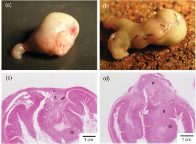
Macroscopic and microscopic observations of the stomach seven days after treatments. Sealing with the PLLA nanosheet (a,c). Conventional suture/ligation (b,d). Letters F, M, and S show fibroblasts, mucosa, and submucosa, respectively. Sealing surgical incisions with this nanosheet instead of performing the traditional suture and ligation procedures is more than just a godsend for Hollywood's plastic surgeons. In cases where internal organs or other tissue has been severely damaged, repair by suture and ligation may become technically difficult and unreliable. Also, internal scarring can lead to sometimes dangerous adhesions, with areas of tissue that should remain separate become joined by scar tissue.Researchers have already been working on novel glue-type (see for instance: Gel-based glue fastens snails to wet surfaces, model for surgical adhesive and sheet-type biomaterials for wound dressing. "Ideally" explains Takeoka, "desirable properties of a novel wound dressing material should incorporate the advantages of both types, that is, not only sufficient adhesive strength to cover the wound site as with a sheet-type structure, but also compatibility with the site as with a glue-type structure."The nanosheet developed by the Japanese team fits that bill – it has very interesting features such as high adhesiveness, high flexibility, and high transparency at the same time. PLLA and other polyesters have been clinically applied in drug delivery and as degradable stitches before, but not as nano-thin sheets. Interestingly, the mechanical properties of the nanosheets are very different from the conventional thin film of the same components.

t
Preparation of PLLA nanosheets and appearance
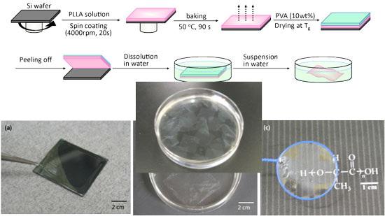
Preparation of PLLA nanosheets and appearance. (Image: Dr. Shinji Takeoka, Waseda University)

Takeoka explains that his and his team's scientific backgrounds are bio-related macromolecular chemistry and molecular assembling science and engineering." We have studied platelet substitutes, which are biodegradable nanoparticles such as phospholipid vesicles and polymerized albumin particles carrying platelet membrane proteins and peptides for ten years" he says. "We noticed that the nanoparticles specifically accumulated at sites of vascular injury and had hemostatic ability. Moreover, in order to enhance their hemostatic abilities, we proposed the idea of sheet-shaped carriers having a larger contact area for a targeting site than spherical nanoparticles." To demonstrate the suitability of their nanosheets as a novel wound dressing material that lacks adhesive reagents, the researchers repaired incisions on mouse stomachs with them instead of sutures. They found that the PLLA nanosheet has an excellent sealing efficacy and the healed wound showed neither scar nor tissue adhesion. Although it will be several years before this material is developed enough to enter clinical trials, Takeoka expects that the nanosheet will be applied not only in the field of surgery as a wound dressing instead of conventional suturing operation but also in the field of plastic surgery, endoscopic surgery, regeneration medicine, and external use (skin etc.). He also sees applications in the fields of material science, material engineering, and surface science.